Spectroscopy Basics
When light of a fixed wavelength impinges upon a material, it may be absorbed and re-emitted by the material as a spectrum of wavelengths of different intensities. Spectral features result from the transition of electrons among the energy levels of the material, and thus provide quantum mechanical information about the material’s structure and composition. The study of emitted light can provide a powerful characterization method to discover new materials and improve old ones. It can allow a geologist to discover information about the mineral composition of a rock sample, and allow a chemist to determine the chemical composition of a sample.
Spectroscopy is broken down into several categories depending on how light interacts with the material of interest. Some common types of spectroscopy are photoluminescence, Raman, and reflectance. These spectroscopic methods are non-destructive and provide detailed insight, at the quantum level, into the composition and characteristics of the material without having to break, etch, machine, or otherwise destroy the sample. An instrument capable of providing a suite of such measurements to a scientist or engineer can enable major discoveries of new materials, but it can also allow users to get a near-instant answer to the question of whether what they grew, spun, pulled, sputtered, or otherwise produced is what they think it is. Spectroscopic instruments with multiple functions are as valuable to a materials researcher as an oscilloscope is to an electrical engineer.
PL: Photoluminescence
Photoluminescence (PL) is a contactless, non-destructive, versatile and powerful optical method to probe the electronic structure of materials. PL occurs when light is absorbed and re-emitted at a range of longer wavelengths. (In chemistry and biology this is typically referred to this as fluorescence).
A monochromatic source of light, typically a laser, is used to excite the sample. The laser light incident on the sample excites electrons from the ground state to a higher energy excited state. The material then releases this energy as a combination of phonons (vibrations) and photons (light) as it relaxes back to the ground state. This light can be collected and analyzed spectrally and spatially to yield important information about the material’s properties. In the figure, the excited states are in the conduction band. The excitation photon should be shown exciting the electron high into the CB. Then non-radiative relaxation brings the electron to the conduction-band minimum (CBM).
Typically, the emitted light is of lower energy than is used for excitation, since some energy is lost to non-radiative processes such as phonon generation. The energy of the emitted light is related to the difference between the two energy levels involved in the transition between an excited state and an equilibrium state.
Fluorescence and PL are used extensively in biology and the study of the dynamics of living cells, where fluorescent, molecular tags are attached to molecules of interest and illuminated by white or monochromatic light. The fluorescence by the tags yields both spatial and compositional information for the investigator. The same phenomena are used in devices such as light emitting diodes and fluorescent lamps. Light is ideal as an investigative tool to discover the electronic properties of common and exotic substances.
Typical applications include:
Defect detection and impurity levels
Recombination mechanisms
Material quality
Band gap determination
Molecular structure and crystallinity
Investigators of semiconductor materials generally select an incident wavelength corresponding to a photon energy greater than the optical bandgap. The figure below shows several compound semiconductors and their bandgaps. An investigator interested in GaN necessarily requires a PL source in the UV while one interested in AlGaAs quantum wells can get by with a longer-wavelength source. An instrument providing automated collection of spectra across a sample with a simple, easily reconfigured optical addressing setup, would provide its owner with the equivalent of an optical ‘Swiss Army knife’ providing multiple measurement capability from a single, compact tool. Klar Scientific’s line of Mini-Pro spectroscopic mapping microscopes allow a user to easily reconfigure it to his/her needs using Wavelength Module Kits, all of which are easily swapped and affordable.
Raman
Raman spectroscopy is a method of accessing information about the molecular vibration in a compound, such as the vibrational modes (phonons) of a semiconductor. Raman signals occur due to the change in polarizability of a molecule. Raman scattering is thus a measure of how much a particular covalent bond deforms in an electric field (from the incident light).
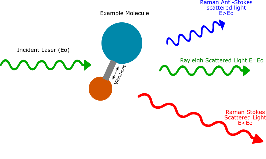
The Raman scattering process is the inelastic scattering of light from molecules in the sample. Incoming photons can lose energy by exciting a vibrational mode (Stokes) or gain thermal energy from molecular vibrations (anti-Stokes). The Raman shift does not depend on the frequency of incident light, but the intensity of the signal can be affected by the choice of excitation wavelength.
To understand this better, imagine the material is a bunch of bowling balls connected by springs. Now imagine the photons of light are marbles, and are launched with a known energy at the bowling ball and spring lattice structure. Most of them will just bounce off without loss of energy, a process known as elastic Rayleigh scattering. However, a few will lose energy and make the bowling ball vibrate. If we measure the energy loss, we get a Stokes shift. It’s also possible for a small vibration in the lattice to give energy to the marble, resulting in a marble with higher energy.
Large libraries of Raman signatures from various materials are now available. For this reason, Raman spectroscopy is often used for molecular ‘fingerprinting’ to identify a particular compound. It is also used extensively in biology and chemistry for drug identification. Polymers have many complex bonds that can vibrate in a variety of ways. Different phases of the same material will have different bond properties, so Raman can be used to give information on the phase of the material. Stress and strain in a material deforms the molecular bonds, causing them to vibrate at a slightly different frequency. In this way, small shifts in a Raman line can show strain in a material.
Excellent but expensive Raman spectroscopy instruments are available in the marketplace. Klar’s instruments target scientists and engineers with more modest budgets whose primary interest is in PL but who occasionally need to make a Raman measurement as well. With Klar’s interchangeable wavelength module kits, a user can perform PL measurements at a variety of excitation wavelengths. Klar also offers a Raman Spectroscopy kit at 532 nm, which includes a more powerful laser and more sensitive spectrometer. For users whose needs span multiple types of spectroscopy, Klar’s reconfigurable, modular instruments are an ideal, cost-effective solution.


